ICON Eyecare Glossary of Eye Terms
There are common eye terms and some not so common eye terms that patients may hear when they visit an eye doctor. At ICON Eyecare in Grand Junction, we encourage you to ask any questions that you may have about your eyecare. We have also prepared a glossary of eye terms you can refer to when you have questions about vision health.
Our team of eye doctors has put together this glossary of items for easy reference. Remember, we’re just a phone call away if you need more information about your eye condition and care.
Age-related macular degeneration: This vision impairment develops when the central part of the retina deteriorates. Millions of Americans develop this condition each year. Although there is no known cure for age-related macular degeneration, treatments can slow the condition. An accurate diagnosis often requires in office testing. It’s more common in females and becomes more common in patients aged 60 and older.
American Optometric Association: Founded in 1898, the AOA (American Optometric Association) is the leading authority on quality eye care in the U.S. and represents more than 44,000 optometry students, optometric professionals, and optometry doctors.
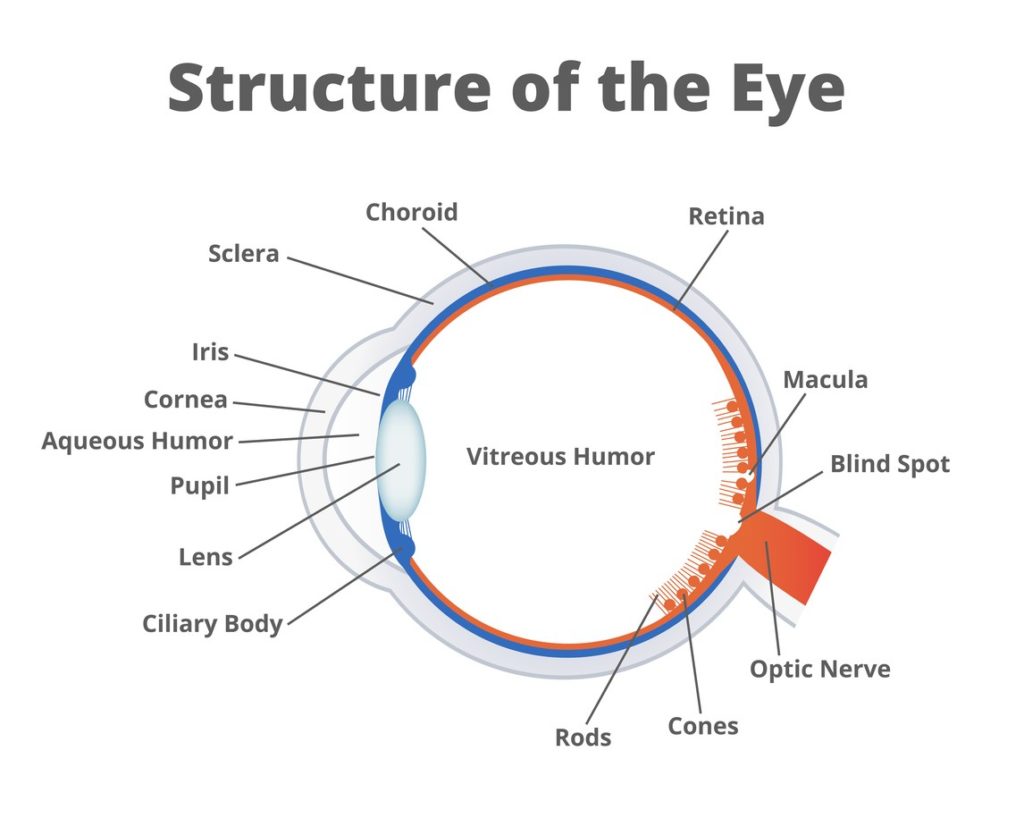
Aqueous humor: This is the watery clear fluid between the eye’s cornea and lens.
Binocular vision: The coordinated use of two eyes to produce a single three-dimensional image. Binocular vision overlaps the visual field of both eyes to avoid blind spots.
Blind spot: When the visual field is tested, this is the blind area that corresponds to the optic disk without any light-sensitive cells where the optic nerve fibers exit the eye. A portion of the visual field remains hidden. However, when both eyes focus on an object, the fields of vision overlap to overcome individual blind spots in the left and right eye. When one eye is closed, blind spots become more apparent.
Blurry vision: Blurry vision is a common eye problem where the vision is no longer clear and in focus. Sudden blurred vision can be caused by any of the eye’s components, including the optic nerve, retina, or cornea. Long-term medical conditions normally cause slowly progressive blurred vision, while a single event often causes sudden blurring. It’s important to visit an eye doctor in Grand Junction if blurry vision does not clear or becomes worse.
Bowman’s membrane: The corneal layer between the stroma and the epithelium made of collagen fibers. Bowman’s membrane supports the cornea.
Bright light: The eye’s main defense against bright light is the iris. This colored part of the eye reduces and enlarges the pupil’s size. When intense light rays hit the eye, the iris constricts the pupil, and this protects the retina and helps it process the image better. Intense light can damage the retina and other sensitive parts of the eye responsible for focusing light rays and creating images.
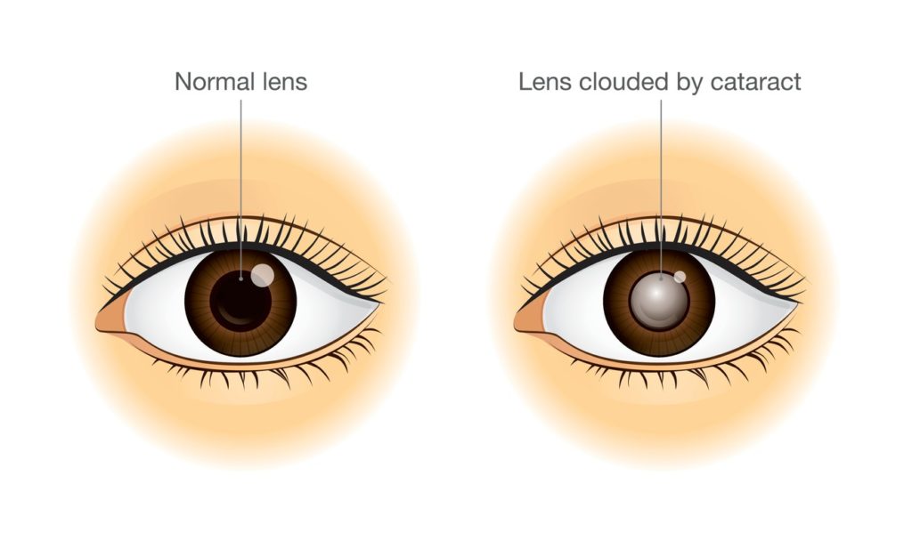
Cataract surgery: With cataract surgery, the lens of the eye is removed and replaced with an artificial lens. The eye’s lens is normally clear, but a cataract makes the lens cloudy, which eventually affects vision. In the early stages, cataracts may not affect vision. However, once cataracts begin to impair vision, it’s a good idea to seek treatment, including surgical or nonsurgical treatment options.
Child’s eye: Many patients wonder when to bring a child in for their first eye exam. We recommend starting as early as age four or five. however, children with poor vision may need eyeglasses or other treatment as early as age two or sooner.
Color blindness: In short, color blindness means that someone cannot distinguish certain colors. Inherited color blindness includes blue (tritan), green (deutan), and red (protan) color vision deficits. These impairments occur in varying degrees of severity. Loss of the blue receptor (tritanopia) is inherited in an autosomal recessive fashion, while green and red and color vision defects are X-linked. So, you can inherit color blindness in multiple ways. Fortunately, there are treatment options for some kinds of color blindness as well as adaptations to help those with color blindness adapt to living with this condition. Unfortunately, there’s no known cure.
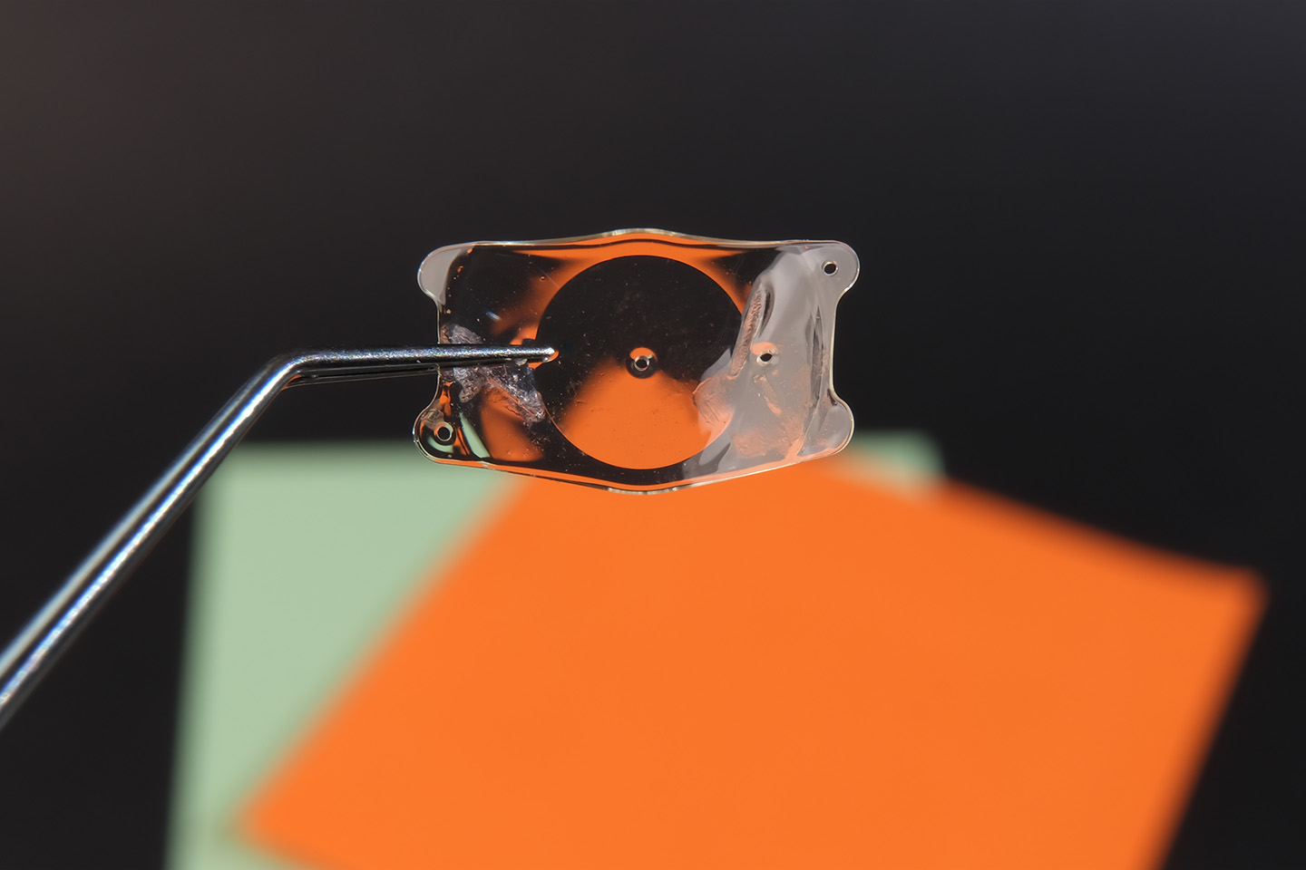
Contact lenses: Contact lenses are prescription lenses that are worn in “contact” with the eyes. They have been designed to maintain ocular health and correct refractive errors, and float on the tear film layer on the cornea’s surface to correct for refractive errors.
Corneal abrasion: This is the loss of the cornea’s epithelial layer, normally caused by minor trauma such as a sports injury, contact lens trauma, and foreign bodies including small bits of metal. Symptoms include foreign body sensation, blurred vision, light sensitivity, grittiness, eye discomfort or pain, a pink or red eye, and tearing.
Corrective lenses: These are lenses worn on or in front of the eyes to correct refractive errors like hyperopia, myopia, presbyopia, and astigmatism. Corrective lenses have been designed to help the eyes properly focus light onto the retina to see clearly.
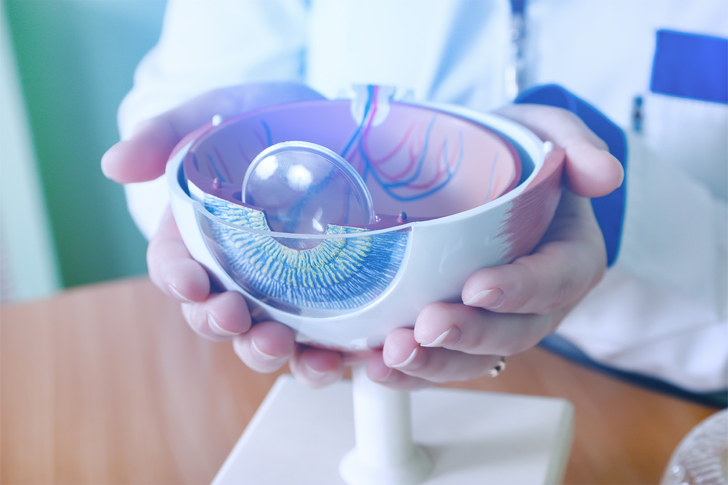
Crystalline lens: The eye’s crystalline lens is the natural lens that produces a third of the eye’s total optical power and focuses light into an image on the retina. This lens is elastic, so it can flex and change its shape.
Depth perception: The blending of two slightly dissimilar images from the eyes to perceive three–dimensional depth.
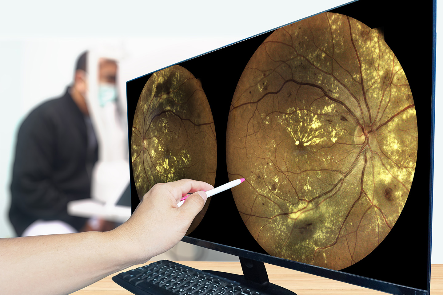
Diabetic retinopathy: Leaking of blood vessels in the retina that is caused by long-term or advanced diabetes which affects the retina and/or macula. There are normally no symptoms at first, but it can result in double vision, blurred near vision, metamorphopsia, retinal/vitreous hemorrhages, and floaters. In later stages, it can also lead to vision loss. Early treatment may include lifestyle changes and treatment from your primary care doctor to prevent worsening of the condition and possible blindness.

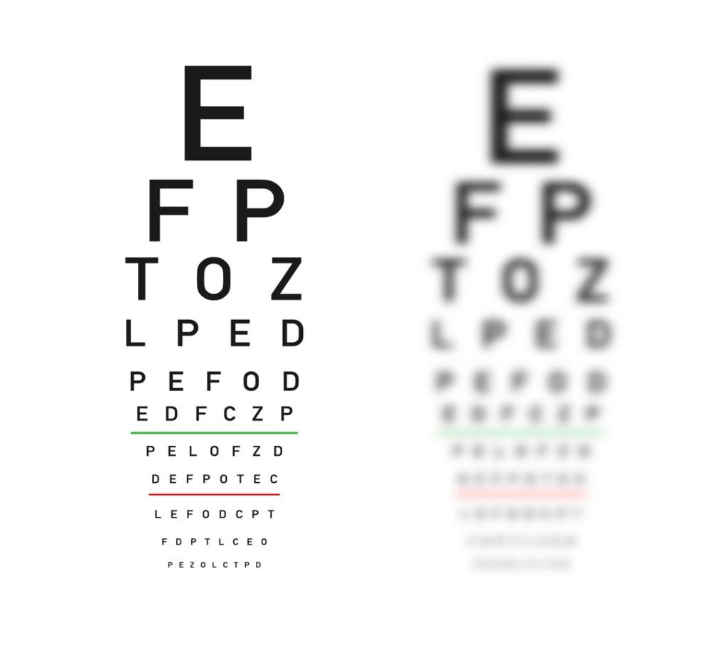
Distant objects: This normally refers to eyesight for objects that are farther away than an arm’s length. Vision for distant objects is required for activities like participating in sports, watching movies and television, and driving. Distance vision is tested using a standard eye chart at about 20 feet away.
Double vision: This is also known as diplopia and occurs when two images of the same object are seen by both or one eye. Double vision can occur after trauma, illness, problems within the brain, or vascular issues like high blood pressure or diabetes. See your eye doctor right away if you experience sudden double vision.
Electrical impulses: Optic nerves send visual information from the retina to the brain. The retina converts the light it receives at the back of the eye into electrical impulses and then sends those impulses to the brain.
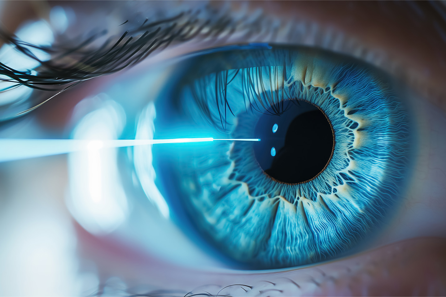
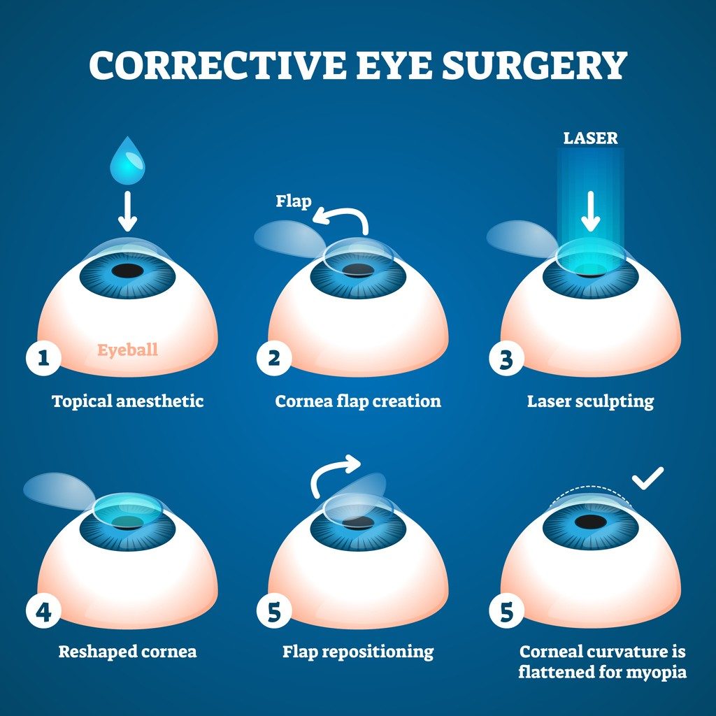
Excimer laser: An instrument that utilizes ultraviolet (shorter wave) light to remove and vaporize tissue from the eye’s surface during a vision correction procedure like PRK and LASIK. Laser surgery allows eye doctors to perform precise, accurate procedures, improving outcomes and shortening recovery time after surgery.
Eye care: Eye care is the treatment and care of vision, eyesight conditions, and eye health in general. It typically includes annual eye exams and treatment for eye conditions due to trauma, injury, illness and other events.
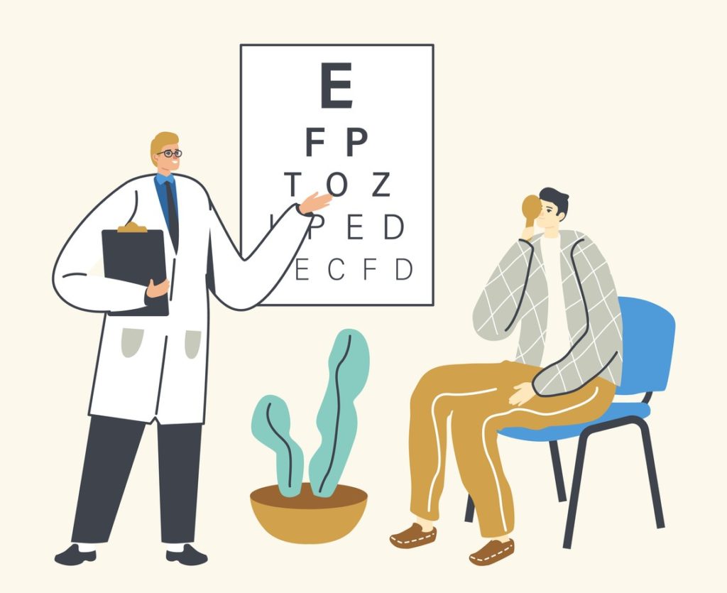
Eye exam: Eye exams involve several tests used to check for eye diseases and evaluate vision. An eye doctor normally uses a variety of instruments, shines bright lights into the eyes, and has their patient look through a variety of lenses. Each test used during eye exams evaluates a different aspect of eye health or vision.
Eye health: Eye health refers to how healthy eyes are. Perfectly healthy eyes provide excellent vision and clear eyes and are free of pain or other symptoms.
Fibrous tissue: Fibrous tissue in the eyes, also known as fibrosis or scarring, can have disastrous consequences for vision. This can result from inflammation or infection in the eye.
Anterior chamber: The anterior chamber at the front part of the eye between the iris and the cornea.
Intraocular lens: This is an artificial lens that is placed in a patient’s eye after the eye’s natural lens has been removed. It has a built-in refractive power designed specifically for a patient’s visual condition, just like a contact lens does.
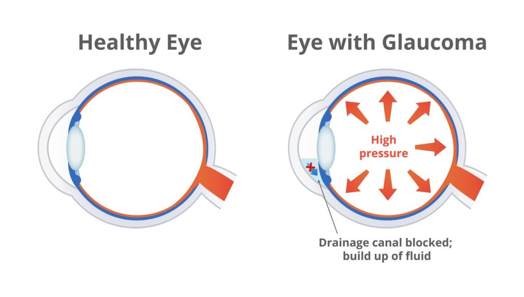
Intraocular pressure: Often abbreviated to IOP, this is eye pressure as determined by the amount of aqueous humor filling the eye. Ocular hypertension (high IOP) can be a sign of glaucoma.
Lazy eye: Lazy eye, also known as amblyopia, is reduced vision in one of the eyes and is caused by abnormal visual development early in life. The lazy, or weaker eye often wanders outward or inward, although it can also appear aligned. Amblyopia normally develops from birth up to 7 years old. It is the main cause of decreased vision among children.
Light rays: When the eye has difficulty focusing light rays onto the retina, it can result in refractive errors such as farsightedness, nearsightedness and astigmatism.
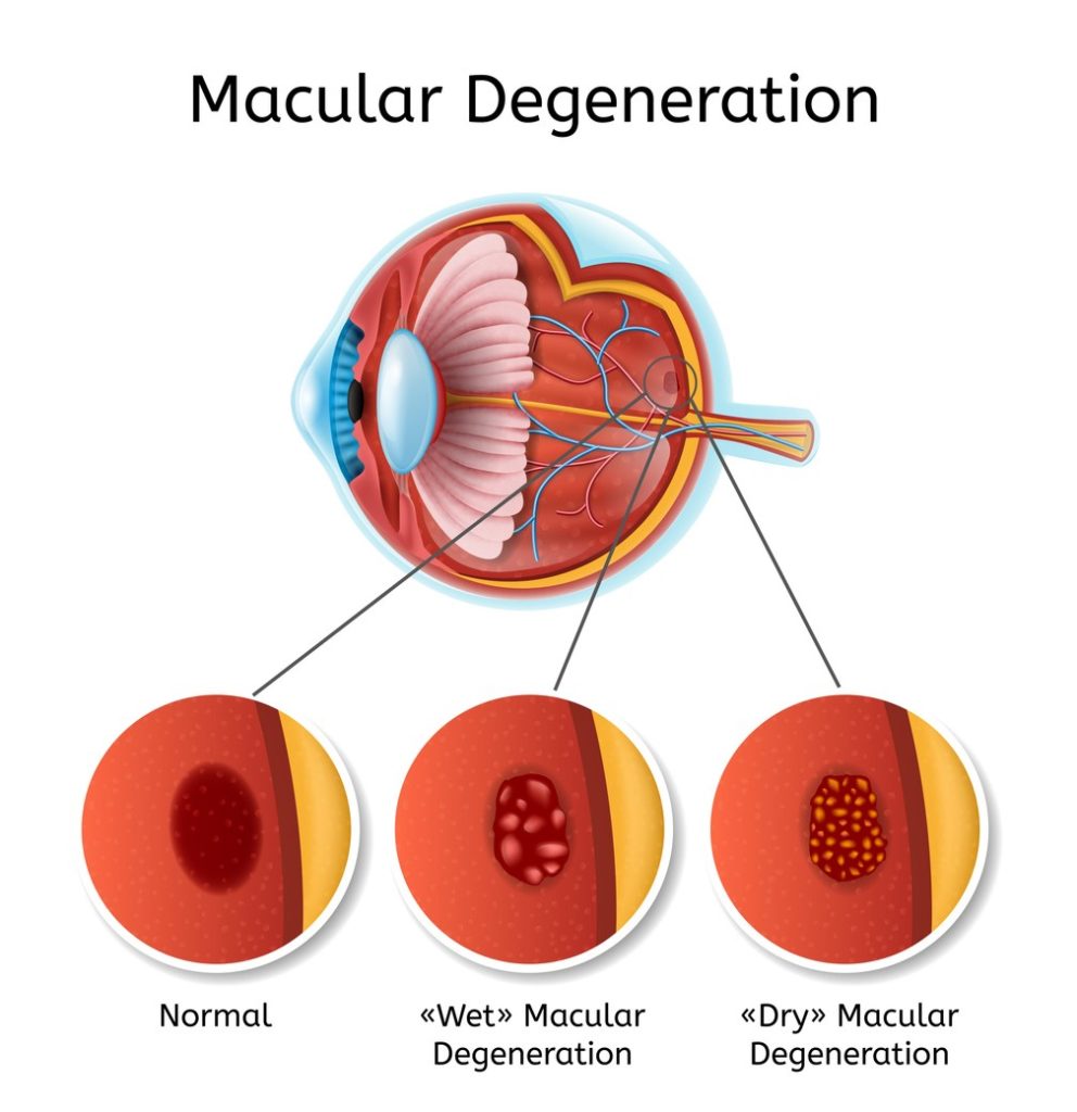
Macular degeneration: This refers to several conditions involving the deterioration of the macula, or center of the retina. It impacts the area where eyesight is normally keenest. This is one of the leading causes of blindness and vision loss in individuals 65 years and older. Macular degeneration is also known as ARMD or AMD (age-related macular degeneration).
Macular edema: Macular edema is the swelling of the macula which makes it difficult to see. It is normally caused by disease or injury.
Optic nerve: This is a bundle of nerves that carries light signals from the retina to the brain, which then converts them into images.
Photorefractive keratectomy: Also referred to as PRK, this is a common alternative to LASIK surgery. If you are not a good candidate for LASIK surgery and have an issue with nearsightedness, this laser surgery may help. The laser changes the shape of the cornea to improve how the eye focuses light on the retina.
Radial keratotomy: Often abbreviated RK, radial keratotomy is a type of refractive surgery done to correct nearsightedness or myopia. It is not a procedure commonly performed nowadays.
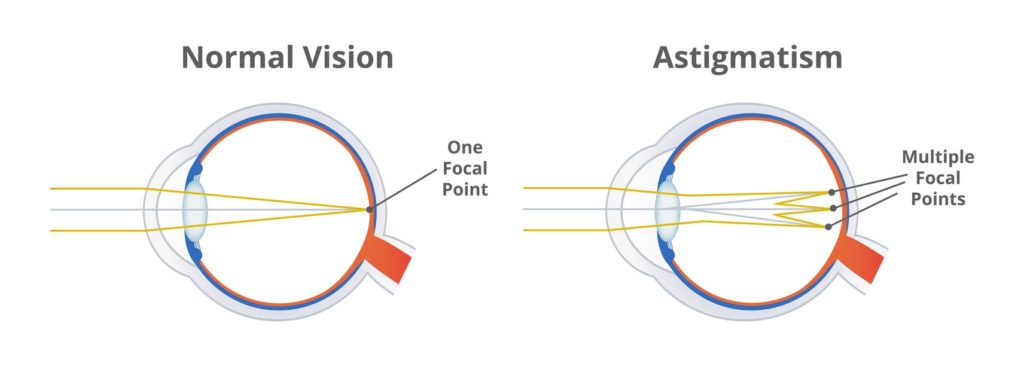
Refractive error: This is when the eyes don’t bend light the way they should, causing images to be out of focus. The most common refractive errors are nearsightedness, farsightedness, and astigmatism. An eye doctor can correct this with contact lenses, prescription glasses, or in some cases with laser corrective surgery.
Refractive surgery: Surgery used to correct visual acuity, with the goal of eliminating or reducing the need for contact lenses or glasses. Includes PRK, radial keratotomy, and LASIK.
Retinal detachment: The retina’s nerve cells normally detect the light that enters an eye and sends signals to the brain about what is seen by the eye. When the retina detaches, you may see a dark curtain in your vision. This is considered an emergency and you should contact your eye doctor immediately as it may result in permanent blindness if not treated promptly.
Retinitis pigmentosa: This is a group of retina conditions that can be inherited. They all cause the loss of sight over time. A decrease in night vision is typically the first sign after which the vision tunnels down to only what is straight ahead. The central vision may eventually also decrease.
Surgical procedure: Any medical procedure that involves an incision with instruments. These are done to arrest disease or repair damage in a living body.
Vision loss: This is losing the ability to see clearly with or without vision correction. Vision correction devices include contact lenses, eyeglasses, surgical correction to the eye, or permanent artificial lenses. Vision loss can occur suddenly or gradually.
Vision problems: Eye diseases such as cataracts, glaucoma, and macular degeneration can cause problems with vision. Symptoms among these disorders vary substantially, so it’s important to have eye exams done regularly. Some vision problems could be dangerous and need to be taken care of immediately.
Visual acuity: This is vision sharpness, usually measured by using a Snellen eye chart. Typically, 20/20 is considered normal vision. It is important to monitor visual acuity over time.
Visual field: The visual field is the total area that can be seen when the eye is pointed forward, including what is seen with peripheral vision.
Vitreous humor: This is the gel-like clear substance located in the center of your eye which helps it maintain shape. Over time, it will pull away from the retina causing a posterior vitreous detachment. If you experience a sudden increase in floaters or flashing lights this is considered urgent and you should call your eye doctor for an evaluation.